14.8
Biological Effects of Ionizing Radiation
Chapter contents: Chapter 14: Nuclear and Particle Physics
We hear many seemingly contradictory things about the biological effects of ionizing radiation. It can cause cancer, burns, and hair loss, yet it is used to treat and even cure cancer. How do we understand these effects? Once again, there is an underlying simplicity in nature, even in complicated biological organisms. All the effects of ionizing radiation on biological tissue can be understood by knowing that ionizing radiation affects molecules within cells, particularly DNA molecules.
Let us take a brief look at molecules within cells and how cells operate. Cells have long, double-helical DNA molecules containing chemical codes called genetic codes that govern the function and processes undertaken by the cell. It is for unraveling the double-helical structure of DNA that James Watson, Francis Crick, and Maurice Wilkins received the Nobel Prize. Damage to DNA consists of breaks in chemical bonds or other changes in the structural features of the DNA chain, leading to changes in the genetic code. In human cells, we can have as many as a million individual instances of damage to DNA per cell per day. It is remarkable that DNA contains codes that check whether the DNA is damaged or can repair itself. It is like an auto check and repair mechanism. This repair ability of DNA is vital for maintaining the integrity of the genetic code and for the normal functioning of the entire organism. It should be constantly active and needs to respond rapidly. The rate of DNA repair depends on various factors such as the cell type and age of the cell. A cell with a damaged ability to repair DNA, which could have been induced by ionizing radiation, can do one of the following:
- The cell can go into an irreversible state of dormancy, known as senescence.
- The cell can initiate programmed cell death.
- The cell can go into unregulated cell division leading to tumors and cancers.
Since ionizing radiation damages the DNA, which is critical in cell reproduction, it has its greatest effect on cells that rapidly reproduce, including most types of cancer. Radiation therapy exploits this sensitivity to kill cancer cells. Targeting the tumor and allowing normal tissue to recover between treatments help limit damage to healthy tissue. Cancer is characterized by a malfunction of cell reproduction, and can also be caused by ionizing radiation. Without contradiction, ionizing radiation can be both a cure and a cause.
To discuss quantitatively the biological effects of ionizing radiation, we need a radiation dose unit that is directly related to those effects. All effects of radiation are assumed to be directly proportional to the amount of ionization produced in the biological organism. The amount of ionization is in turn proportional to the amount of deposited energy. Therefore, we define a radiation dose unit called the rad, as of a joule of ionizing energy deposited per kilogram of tissue, which is
.
(14.8.1)For example, if a 50.0-kg person is exposed to ionizing radiation over her entire body and she absorbs 1.00 J, then her whole-body radiation dose is
.
If the same 1.00 J of ionizing energy were absorbed in her 2.00-kg forearm alone, then the dose to the forearm would be
,
and the unaffected tissue would have a zero rad dose. While calculating radiation doses, you divide the energy absorbed by the mass of affected tissue. You must specify the affected region, such as the whole body or forearm in addition to giving the numerical dose in rads. The SI unit for radiation dose is the gray (Gy), which is defined to be
.
(14.8.2)However, the rad is still commonly used. Although the energy per kilogram in 1 rad is small, it has significant effects since the energy causes ionization. The energy needed for a single ionization is a few eV, or less than . Thus, 0.01 J of ionizing energy can create a huge number of ion pairs and have an effect at the cellular level.
The effects of ionizing radiation depend on the absorbed dose, the type of radiation, and the type of tissue. For a given absorbed dose, densely concentrated ionization, such as that produced by α particles, can cause damage that is harder to repair (see Figure 14.8.1). The relative biological effectiveness (RBE) compares the absorbed doses of a reference radiation and the radiation being studied that produce the same biological effect. It depends on the effect being measured. For radiation protection, standardized radiation weighting factors, , are used instead (see Table 14.8.1). For one type of radiation, the equivalent dose H to an organ or tissue is its average absorbed dose D multiplied by the radiation weighting factor. Absorbed dose is measured in grays (Gy); equivalent dose is measured in sieverts (Sv). The related quality factor is used in dose-equivalent quantities for radiation monitoring; it is not another name for RBE. The older unit roentgen equivalent man (rem) is also used for weighted doses.
(14.8.3)
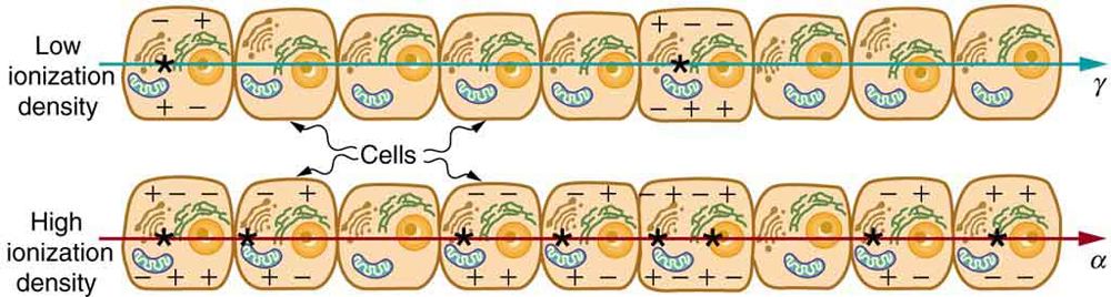
So, if an organ received an absorbed dose of 0.0200 Gy (2.00 rad) of γ radiation, its equivalent dose would be (0.0200 Gy)(1) = 0.0200 Sv (2.00 rem). For the same absorbed dose from α radiation deposited inside the body, the equivalent dose would be (0.0200 Gy)(20) = 0.400 Sv (40.0 rem). The α radiation receives 20 times the weighting for radiation protection; this does not mean that every biological effect is 20 times greater. For a mixture of radiation types, the equivalent doses from each type are added. The sievert (Sv) and rem are related by
.
(14.8.4)The radiation weighting factors in Table 14.8.1 are 1 for β particles, γ rays, and X-rays, three common types of radiation. For these, absorbed dose in Gy and equivalent dose in Sv have the same numerical value, as do absorbed dose in rad and equivalent dose in rem. Neutrons produce ionization indirectly through secondary charged particles, including recoiling nuclei. Their weighting factor depends on neutron energy. To account also for the different sensitivities of organs and tissues, effective dose adds their equivalent doses multiplied by tissue weighting factors. It is also measured in Sv or rem and is used to compare radiation exposures in terms of long-term risk. These weighted doses are protection quantities; absorbed dose in Gy is used to assess immediate tissue injury. Table 14.8.2 summarizes the units used for radiation.
| Type of radiation | |
|---|---|
| X-rays and γ rays | 1 |
| Electrons and positrons (β particles), and muons | 1 |
| Protons and charged pions | 2 |
| α particles, fission fragments, and heavy ions | 20 |
| Neutrons | Depends on neutron energy |
| Quantity | SI unit | Meaning | Older unit | Conversion |
|---|---|---|---|---|
| Activity | becquerel (Bq) | Decays per second | curie (Ci) | 1 Bq ≈ 2.7 × 10⁻¹¹ Ci |
| Absorbed dose | gray (Gy) | Energy deposited per mass; 1 Gy = 1 J/kg | rad | 1 Gy = 100 rad |
| Equivalent dose | sievert (Sv) | Absorbed dose weighted for radiation type | rem | 1 Sv = 100 rem |
| Effective dose | sievert (Sv) | Equivalent doses weighted for tissues and added | rem | 1 Sv = 100 rem |
Misconception Alert: Activity vs. Dose
“Activity” refers to the radioactive source while “dose” refers to the amount of energy from the radiation that is deposited in a person or object. A high level of activity doesn’t mean much danger if a person is far away from the source, or if substantial shielding is placed between the source and the person. A radioactive source can have a high activity in two different ways—the type of radioactive isotope may have a short half-life, resulting in more number of decays per second for a given amount of material, or there may be more material, resulting in more total number of decays per second.
For a given radioactive source with given activity, the dose received by nearby personnel can be reduced by radiation safety practices of establishing minimum distance perimeters (intensity of radiation follows inverse-square law) and designing and placing shielding materials around the source.
The large-scale effects of radiation on humans include acute radiation sickness and long-term effects. Table 14.8.3 gives the effects of large whole-body doses received in a short time. These effects may develop over days or weeks. If the radiation exposure is spread out over more time, greater doses are needed to cause the effects listed. This is due to the body’s ability to partially repair the damage. Exposure of a small part of the body can cause local tissue injury without whole-body radiation sickness. For X-rays and gamma rays, an absorbed dose less than 0.1 Gy (10 rad) is called a low dose, 0.1 to 1 Gy (10 to 100 rad) a moderate dose, and greater than 1 Gy (100 rad) a high dose.
| Absorbed dose (Gy) | Major effects |
|---|---|
| About 0.7–10 | Bone-marrow damage reduces blood cell production, increasing the risk of infection and bleeding. |
| Above about 10 | Severe intestinal damage, in addition to bone-marrow damage; survival is unlikely. |
| Above about 50 | Severe damage to the circulatory and central nervous systems; rapidly fatal. |
Immediate effects are explained by the effects of radiation on cells and the sensitivity of rapidly reproducing cells to radiation. The first clue that a person has been exposed to radiation is a change in blood count, which is not surprising since blood cells are the most rapidly reproducing cells in the body. At higher doses, nausea and hair loss are observed, which may be due to interference with cell reproduction. Cells in the lining of the digestive system also rapidly reproduce, and their destruction causes nausea. When the growth of hair cells slows, the hair follicles become thin and break off. High doses cause significant cell death in all systems, but the lowest doses that cause fatalities do so by weakening the immune system through the loss of white blood cells.
Radiation can cause cancer and can damage genetic material. Damage to reproductive cells can potentially be passed to offspring. For high doses of radiation, the risk of cancer is reasonably well known from studies of exposed groups. Hiroshima and Nagasaki survivors and a smaller number of people exposed by their occupation, such as radium dial painters, have been fully documented. Chernobyl victims will be studied for many decades, with some data already available. For example, a significant increase in childhood thyroid cancer has been observed. The risk of a radiation-induced cancer for low and moderate doses is generally assumed to be proportional to the risk known for high doses. Under this assumption, any dose of radiation, no matter how small, involves a risk to human health. This is called the linear hypothesis, or linear no-threshold model, and is used in radiation protection. The size of the cancer risk at very low doses remains uncertain. Cells can repair radiation damage, but some damage may remain or be repaired incorrectly.
Radiation-induced cancers can appear years after exposure. Among Hiroshima and Nagasaki survivors, leukemia began to increase about 2–3 years after exposure, and solid cancers about 10 years after exposure. Increased cancer risk can persist for decades. Omitting many details, the additional lifetime risk of dying from radiation-induced cancer is about in per rem of whole-body exposure, within an order of magnitude.
If a person receives a whole-body dose of 1 rem, this estimate gives an additional lifetime risk of dying from radiation-induced cancer of about 300 in a million, or 0.03 percent. Since cancer is already a common cause of death, such a small increase is difficult to detect statistically. For Hiroshima and Nagasaki survivors within 2,500 m of the hypocenters, the reported average dose was about 0.2 Gy, roughly comparable to 20 rem of whole-body gamma exposure. Our linear estimate would give an additional lifetime risk of about 0.6 percent for that exposure. Both risk estimates are approximate, within an order of magnitude. The Radiation Effects Research Foundation’s Life Span Study has detected increased cancer mortality among Hiroshima and Nagasaki survivors. For comparison, RERF gives a background lifetime cancer mortality risk of roughly 20 percent for unexposed people.
The risk of heritable effects from radiation is poorly known in humans. For example, there is no evidence of increased genetic defects among the offspring of Hiroshima and Nagasaki survivors. Animal studies help estimate this risk. For radiation protection, the linear hypothesis is used to estimate risks at low doses. Hormesis is a term used to describe generally favorable biological responses to low exposures of toxins or radiation, but a health benefit from low-dose radiation has not been established for humans.
Even the linear hypothesis estimates of the risks are relatively small, and the average person is not exposed to large amounts of radiation. Table 14.8.4 lists global average annual effective doses from natural background radiation, totaling about 3 mSv (300 mrem). Cosmic rays are partially shielded by the atmosphere, and the dose depends upon altitude and latitude. Radon exposure also varies with local geology and the construction and ventilation of buildings.
Doses from the Earth itself are mainly due to the isotopes of uranium, thorium, and potassium, and vary greatly by location. Some places have great natural concentrations of uranium and thorium, yielding much higher terrestrial radiation doses—for example, areas with thorium-rich sands along the Kerala coast in India. Internal doses come from naturally occurring radioactive isotopes in foods and liquids that we ingest, including potassium-40 and carbon-14. So we are all a little radioactive. Medical and dental diagnostic exposures are mostly from X-rays. It should be noted that X-ray doses tend to be localized and are becoming much smaller with improved techniques. Table 14.8.5 shows typical doses received during various diagnostic X-ray examinations. For comparison, a chest X-ray dose of about 0.1 mSv is comparable to about 12 days of natural background at 3 mSv per year; an abdomen and pelvis CT dose of about 7.7 mSv is comparable to about 2.6 years. These comparisons indicate the scale of effective dose, not the same exposure of each organ. A CT scan collects X-ray measurements from many angles to reconstruct cross-sectional images of the body. These multiple exposures generally result in a higher dose than a conventional X-ray image.
Radon is usually more pronounced underground and in buildings with low air exchange with the outside world. Almost all soil contains some and , but radon is lower in mainly sedimentary soils and higher in granite soils. Thus, the exposure to the public can vary greatly, even within short distances. Radon can diffuse from the soil into homes, especially basements. The estimated exposure for is controversial. Recent studies indicate there is more radon in homes than had been realized, and it is speculated that radon may be responsible for 20 percent of lung cancers, being particularly hazardous to those who also smoke. Many countries have introduced limits on allowable radon concentrations in indoor air, often requiring the measurement of radon concentrations in a house prior to its sale. Ironically, it could be argued that the higher levels of radon exposure and their geographic variability, taken with the lack of demographic evidence of any effects, means that low-level radiation is less dangerous than previously thought.
Radiation Protection
Laws regulate radiation doses to which people can be exposed. Dose limits depend upon the country and the type of exposure. For example, the 2025 U.S. Nuclear Regulatory Commission regulations set an annual occupational limit of 50 mSv in total effective dose equivalent for adults. Higher local doses are allowed for the hands. A lower limit applies to the embryo or fetus of a worker who has declared her pregnancy in writing. For members of the public, the general annual limit from licensed operations is 1 mSv, excluding background radiation and doses received as a medical patient, among other exclusions. Radiation protection follows the ALARA principle: keep doses as low as reasonably achievable, even when they are below regulatory limits. Radiation detectors are used to monitor exposure and evaluate protective measures.
| Source | Average annual effective dose (mSv) |
|---|---|
| Inhalation, mainly radon, thoron, and their decay products | 1.8 |
| Cosmic radiation | 0.3 |
| External radiation from the ground and building materials | 0.4 |
| Ingestion in food and water | 0.5 |
| Total | 3.0 |
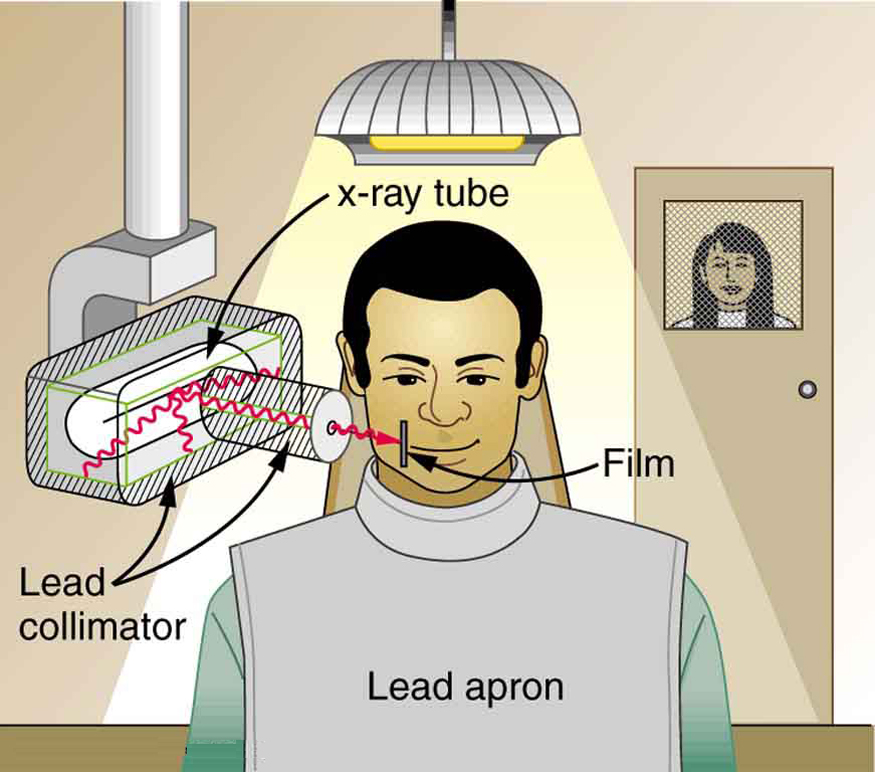
To physically limit radiation doses from external sources, we limit the time of exposure, increase the distance from a source, and use shielding.
Figure 14.8.2 illustrates how these are used to protect both the patient and the dental technician when an X-ray is taken. The less time a person is exposed to a given source, the smaller is the dose received by the person. The greater the distance from the source, the more the radiation spreads out. Shielding absorbs radiation, and the appropriate type of shielding depends on the type of radiation.
| Procedure | Approximate effective dose (mSv) |
|---|---|
| Chest X-ray | 0.1 |
| Dental panoramic X-ray | 0.025 |
| Skull X-ray | 0.07 |
| Limb or joint X-ray (except hip) | < 0.01 |
| Screening digital mammogram | 0.28 |
| Barium enema | 6 |
| Upper GI study with barium | 6 |
| CT brain | 1.6 |
| CT abdomen and pelvis | 7.7 |
Risk versus Benefit
Medical doses of radiation are also limited. Diagnostic doses are generally low and have been further lowered with improved techniques and more efficient detectors. Radiation should be used diagnostically only when needed so that the risk is justified by the benefit of the diagnosis. Chest X-rays give low doses—about 0.1 mSv effective dose for an adult. Other X-ray procedures give higher doses, such as about 6 mSv in a chest CT scan. Dental X-ray doses depend on the type and number of images; a panoramic image of the whole mouth gives an effective dose of about 0.025 mSv. These are approximate values that vary with the patient and the technique used. Medical images with radiopharmaceuticals also involve radiation exposure. One example is the thyroid scan using , which has a relatively long half-life of about 8 days and emits beta particles as well as gamma rays. The isotope has a shorter half-life of about 13 hours and does not emit beta-minus particles, which can reduce thyroid exposure in diagnostic imaging. The dose depends on the activity administered and how much of the isotope the thyroid takes up.