14.2
Nuclear Radioactivity
Chapter contents: Chapter 14: Nuclear and Particle Physics
The discovery and study of nuclear radioactivity quickly revealed evidence of revolutionary new physics. In addition, uses for nuclear radiation also emerged quickly—for example, people such as Ernest Rutherford used it to determine the size of the nucleus and devices were painted with radium-doped paint to make them glow in the dark (see Figure 14.2.1). We therefore begin our study of nuclear physics with the discovery and basic features of nuclear radioactivity.

Discovery of Nuclear Radioactivity
In 1896, the French physicist Antoine Henri Becquerel (1852–1908) accidentally found that a uranium salt emits invisible, penetrating rays that can darken a photographic plate enclosed in an opaque envelope. The rays therefore carry energy; but amazingly, the salt emits them continuously without any energy input. This is an apparent violation of the law of conservation of energy, one that we now understand is due to the conversion of a small amount of mass into energy, as related in Einstein’s famous equation . It was soon evident that Becquerel’s rays originate in the nuclei of the atoms and have other unique characteristics. The spontaneous transformation of unstable nuclei is called nuclear radioactivity or simply radioactivity. The rays themselves are called nuclear radiation. An unstable nucleus that spontaneously transforms into another nucleus or a lower-energy nuclear state is said to decay (a term also used to describe the emission of radiation by atoms in excited states). A substance or object that emits nuclear radiation is said to be radioactive.
Two types of experimental evidence imply that Becquerel’s rays originate not from the atomic structure, but something smaller inside. First, the radiation is found to be associated with certain elements, such as uranium. Radiation does not vary with chemical state—that is, uranium is radioactive whether it is in the form of an element or compound. In addition, radiation does not vary with temperature, pressure, or ionization state of the uranium atom. Since all of these factors affect electrons in an atom, the radiation cannot come from electron transitions, as atomic spectra do. The huge energy emitted during each event is the second piece of evidence that the radiation cannot be atomic in origin. Nuclear radiation has energies of the order of per event, which is much greater than the typical atomic energies (a few ), such as that observed in spectra and chemical reactions, and more than ten times as high as the most energetic characteristic X-rays. Becquerel did not vigorously pursue his discovery for very long. In 1898, Marie Skłodowska-Curie (1867–1934), often known as Marie Curie, began her doctoral study of Becquerel’s rays. She and her husband—French physicist Pierre Curie (1859–1906)—soon discovered two new radioactive elements, which she named polonium (after her native land) and radium (because it radiates). These two new elements filled holes in the periodic table and, further, displayed much higher levels of radioactivity per gram of material than uranium. Over a period of four years, working under poor conditions and spending their own funds, the Curies processed more than a ton of uranium ore to isolate a gram of radium salt. Radium became highly sought after, because it was about two million times as radioactive as uranium. Curie’s radium salt glowed visibly from the radiation that took its toll on them and other unaware researchers. Shortly after completing her Ph.D., both Curies and Becquerel shared the 1903 Nobel Prize in physics for their work on radioactivity. Pierre was killed in a horse cart accident in 1906, but Marie continued her study of radioactivity for nearly 30 more years. Awarded the 1911 Nobel Prize in chemistry for her discovery of two new elements, she remains the only person to win Nobel Prizes in physics and chemistry. Marie’s radioactive fingerprints on some pages of her notebooks can still expose film, and she suffered from radiation-induced lesions. She died of aplastic anemia likely caused by long-term exposure to radiation, but she was active in research almost until her death in 1934.
Alpha, Beta, and Gamma
Research begun by people such as New Zealander Ernest Rutherford soon after the discovery of nuclear radiation indicated that different types of rays are emitted. Eventually, three types were distinguished and named alpha (), beta (), and gamma (), because, like X-rays, their identities were initially unknown. Figure 14.2.2 shows what happens if the rays are passed through a magnetic field. The ’s are unaffected, while the ’s and ’s are deflected in opposite directions, indicating that the ’s are positive, the ’s negative, and the ’s uncharged. Rutherford used both magnetic and electric fields to show that ’s have a positive charge twice the magnitude of an electron, or . In the process, he found the ’s charge to mass ratio to be several thousand times smaller than the electron’s. Later on, Rutherford collected ’s from a radioactive source and passed an electric discharge through them, obtaining the spectrum of recently discovered helium gas. Among many important discoveries made by Rutherford and his collaborators was the proof that radiation is the emission of a helium nucleus. Rutherford won the Nobel Prize in chemistry in 1908 for his early work. He continued to make important contributions until his death in 1937.
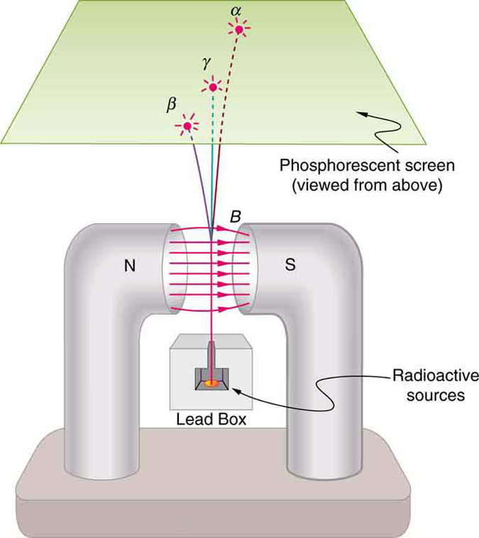
Other researchers had already proved that ’s are negative and have the same mass and same charge-to-mass ratio as the recently discovered electron. By 1902, it was recognized that radiation is the emission of an electron. Although ’s are electrons, they do not exist in the nucleus before it decays and are not ejected atomic electrons—the electron is created in the nucleus at the instant of decay.
Since ’s remain unaffected by electric and magnetic fields, it is natural to think they might be photons. Evidence for this grew, but it was not until 1914 that this was proved by Rutherford and collaborators. By scattering radiation from a crystal and observing interference, they demonstrated that radiation is the emission of a high-energy photon by a nucleus. In fact, radiation comes from the de-excitation of a nucleus, just as an X-ray comes from the de-excitation of an atom. The names “ ray” and “X-ray” identify the source of the radiation. At the same energy, rays and X-rays are otherwise identical.
| Type of Radiation | Typical penetration and shielding |
|---|---|
| Stopped by a sheet of paper; travels a few cm in air or fractions of a mm in tissue | |
| Stopped by a thin layer of plastic or aluminum; typically penetrates a few mm of tissue, depending on energy | |
| Several cm of lead or meters of concrete can substantially reduce intensity; the required thickness depends on photon energy, source strength, and the reduction needed |
Ionization and Range
Two of the most important characteristics of , , and rays were recognized very early. All three types of nuclear radiation produce ionization in materials, but they penetrate different distances in materials—that is, they have different ranges. Let us examine why they have these characteristics and what are some of the consequences.
Like X-rays, nuclear radiation in the form of ’s, ’s, and ’s has enough energy per event to ionize atoms and molecules in any material. The energy emitted in various nuclear decays ranges from a few to more than , while only a few are needed to produce ionization. The effects of X-rays and nuclear radiation on biological tissues and other materials, such as solid state electronics, are directly related to the ionization they produce. All of them, for example, can damage electronics or kill cancer cells. In addition, methods for detecting X-rays and nuclear radiation are based on ionization, directly or indirectly. All of them can ionize the air between the plates of a capacitor, for example, causing it to discharge. This is the basis of inexpensive personal radiation monitors, such as pictured in Figure 14.2.3. Apart from , , and , there are other forms of nuclear radiation as well, and these also produce ionization with similar effects. We define ionizing radiation as any form of radiation that produces ionization whether nuclear in origin or not, since the effects and detection of the radiation are related to ionization.

The range of radiation is defined to be the distance it can travel through a material. Range is related to several factors, including the energy of the radiation, the material encountered, and the type of radiation (see Figure 14.2.4). For alpha and beta particles, all other factors being the same, the higher the energy, the greater the range. These particles lose energy through many interactions with the material, so a more energetic particle travels farther before stopping. Gamma ray photons are absorbed or scattered in interactions whose probabilities depend on photon energy and the material; they do not have a definite stopping range.
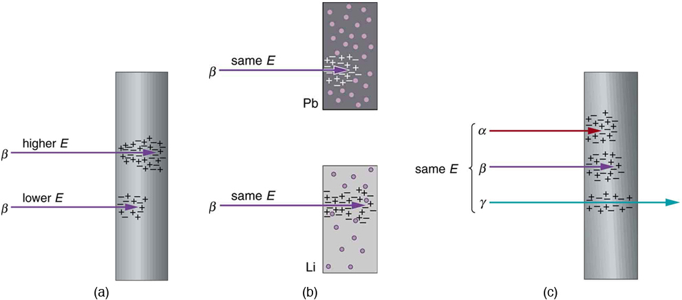
Radiation can be absorbed or shielded by materials. Lead is a particularly effective shield for X-rays and gamma rays compared with materials such as plastic or air; other types of radiation may require different shielding. How does the range of radiation depend on material? Ionizing radiation interacts best with charged particles in a material. Since electrons have small masses, they most readily absorb the energy of the radiation in collisions. The greater the density of a material and, in particular, the greater the density of electrons within a material, the smaller the range of radiation.
Different types of radiation deposit energy differently. Alpha particles generally produce more concentrated ionization than beta particles of comparable energy in the same material, losing their energy over a shorter distance. Gamma rays produce ionization indirectly, through the charged particles set in motion by their interactions.
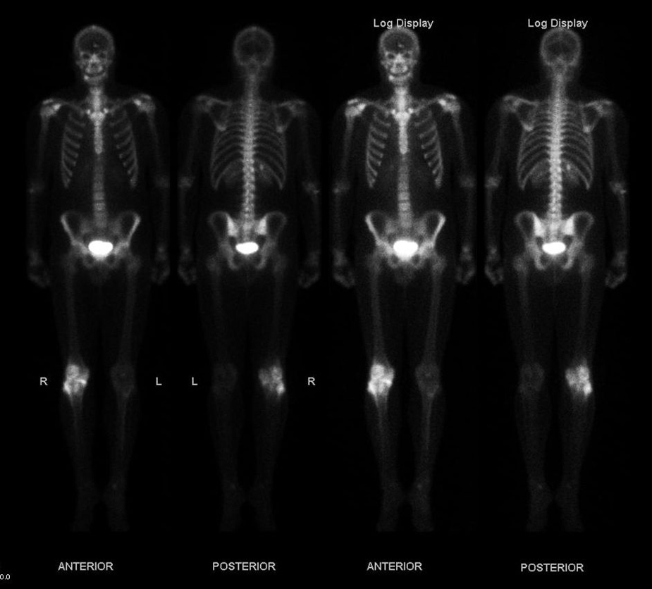
Alpha radiation from radioactive sources has a range much less than a millimeter of biological tissues, usually not enough to even penetrate the dead layers of our skin. On the other hand, the same radiation can penetrate a few centimeters of air, so mere distance from a source prevents radiation from reaching us. This makes radiation less penetrating from outside the body than and radiation. If alpha-emitting material enters the body, it can severely damage nearby tissue. Typical radiation can penetrate a few millimeters of tissue or about a meter of air. Beta radiation is thus hazardous even when not ingested. Beta particles can be stopped by shielding, but their slowing can produce bremsstrahlung X-rays, particularly in materials such as lead. Plastic is often used to stop beta particles while limiting this X-ray production; additional shielding may be needed for the X-rays. Gamma rays do not have a well-defined stopping range, but they are effectively shielded—that is, reduced in intensity to acceptable levels—by sufficient shielding, often many centimeters of lead. The required thickness depends on the photon energy, source strength, and exposure conditions. One benefit of ’s is that they allow radioactive tracers to be detected (see Figure 14.2.5).